
Non-proliferative retinopathy
Damaged vessels may leak or close. Monitoring and systemic risk-factor control are important; the severity determines follow-up.
Screening and treatment for diabetic retinopathy and other retinal conditions—from monitoring and injections to laser and vitreoretinal surgery.
Book a retinal assessmentThe retina is the light-sensitive layer at the back of the eye. Diabetes can damage its small blood vessels, causing leakage, bleeding and reduced blood supply. Early diabetic retinopathy may cause no symptoms, so clear vision does not replace retinal screening.
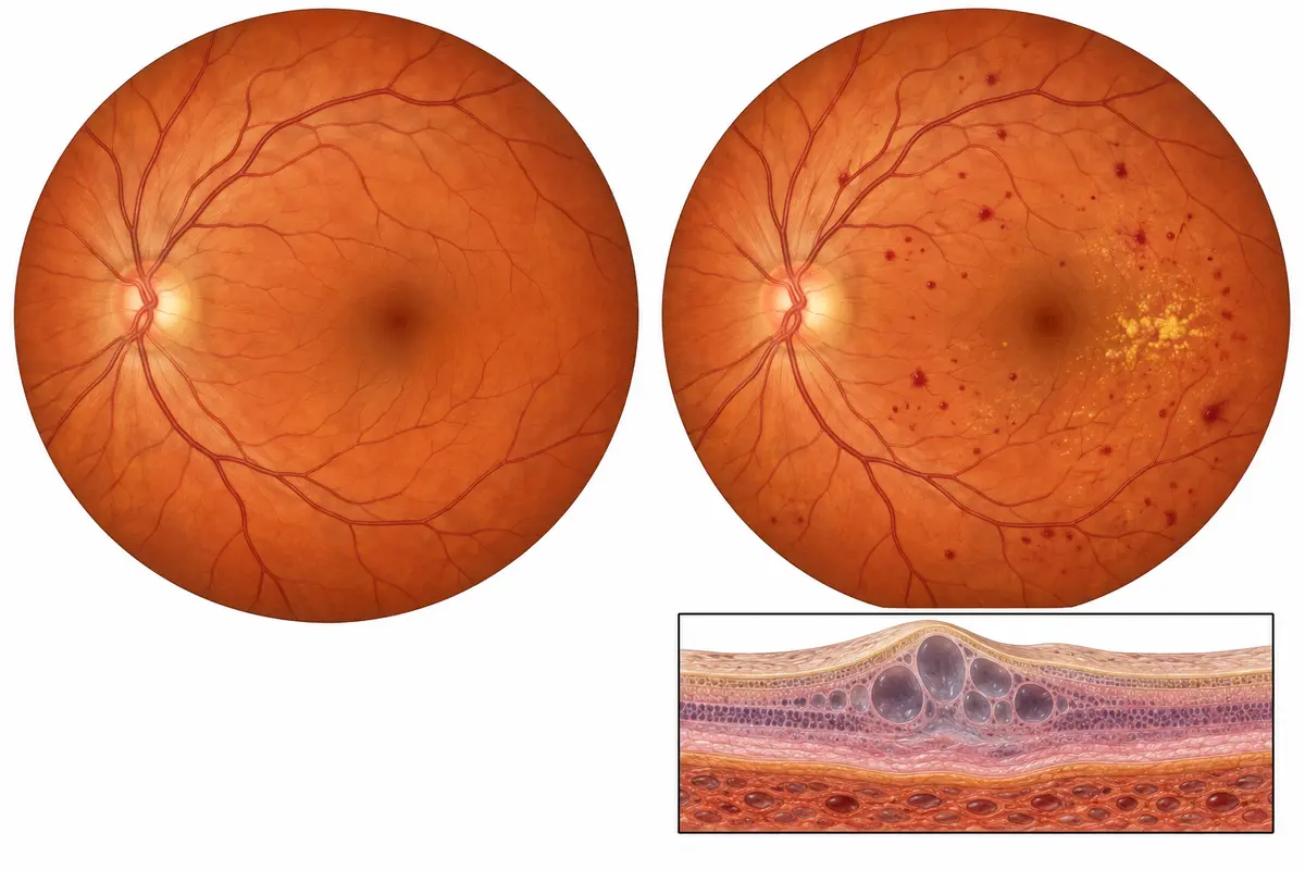
Damaged vessels may leak or close. Monitoring and systemic risk-factor control are important; the severity determines follow-up.
Fluid collects in the macula, the area responsible for detailed central vision. This can affect reading and face recognition. It can occur at different stages of retinopathy.
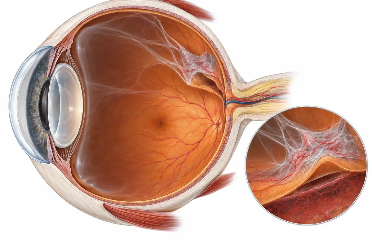
Abnormal new vessels grow in response to poor oxygen supply. They can bleed into the vitreous or develop scar tissue that pulls on the retina.
Assessment includes visual acuity and a dilated retinal examination. OCT measures macular swelling and structural changes. Retinal photography documents disease; fluorescein angiography or OCT angiography may clarify leakage or blood flow when needed. Ultrasound can help when bleeding blocks the view. Treatment depends on these findings, not on symptoms alone.
Bring your medication list, recent HbA1c results and previous eye scans. Dilation can temporarily blur vision and increase light sensitivity; arrange transport and avoid driving until vision has recovered.
Glucose targets depend on factors including age, other illnesses and the risk of hypoglycemia. Coordination with diabetes care is especially important during major treatment changes and pregnancy.
| Treatment | Main role | Key point |
|---|---|---|
| Monitoring | Track early or stable disease | Repeat examination and scans detect change before symptoms develop. |
| Intravitreal injections | Reduce macular swelling or abnormal vessel activity | Repeated treatment and OCT review are often needed. |
| Retinal laser | Treat proliferative disease or selected leakage | It protects vision; it does not guarantee recovery of lost sight. |
| Vitrectomy | Remove persistent blood or relieve retinal traction | Used for selected advanced disease and retinal detachments. |
These are possible pathways, not a fixed sequence. More than one treatment may be needed.
Anti-VEGF medicines reduce leakage and abnormal vessel activity. They are commonly used for vision-impairing diabetic macular edema and selected proliferative disease. Treatment often begins with frequent injections; later intervals depend on response. Steroid injections or implants may suit selected cases, but can raise eye pressure and increase cataract risk.
The eye is numbed and cleaned, then medication is placed into the vitreous through the white of the eye. Brief pressure or irritation can occur. Rare complications include serious infection and retinal detachment. Increasing pain or deteriorating vision after treatment is urgent.
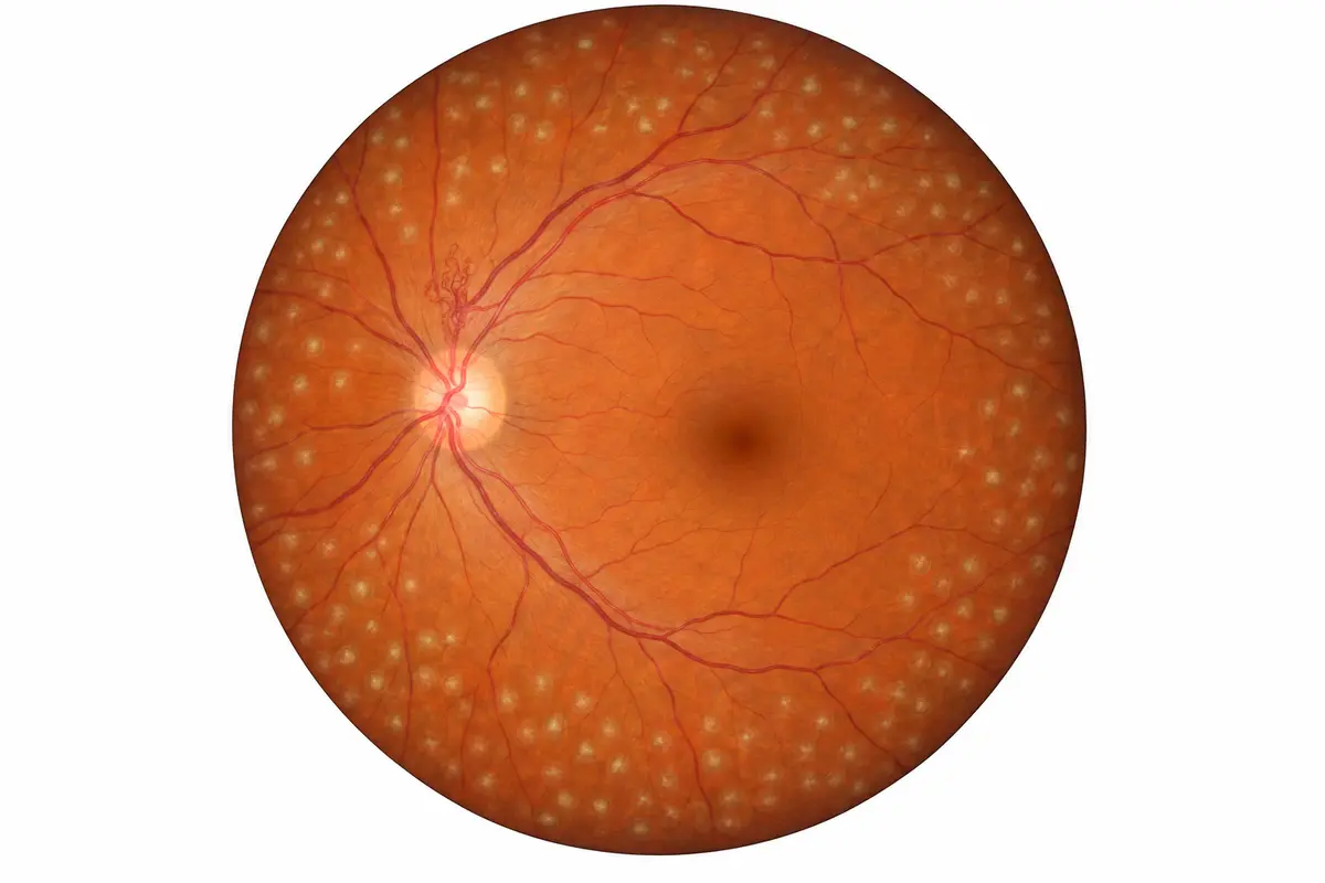
Panretinal photocoagulation (PRP) treats areas of peripheral retina to reduce the drive for abnormal new vessels in proliferative disease. Focal or grid laser may address selected areas of leakage. Laser and injections sometimes form part of the same plan.
More than one session may be needed. PRP can affect peripheral and night vision. Its main benefit is reducing the risk of further severe sight loss.
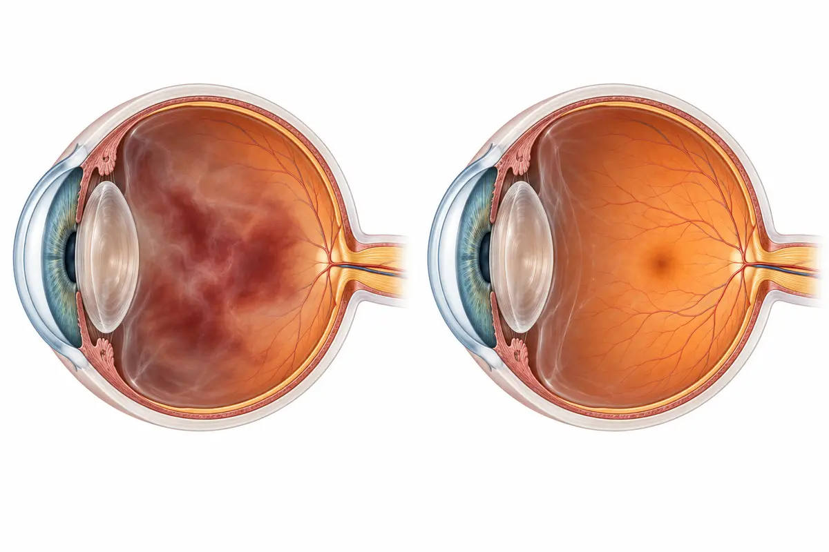
Vitrectomy removes the vitreous gel and blood that prevents light reaching the retina. Scar tissue can be released when it pulls on the retina; laser may be applied during surgery. Persistent vitreous hemorrhage and tractional retinal detachment are reasons surgery may be needed.
Gas or silicone oil may support the retina in selected operations. If gas is present, flying and high-altitude travel are unsafe until it has gone, and nitrous oxide anesthesia must be avoided. Positioning depends on the operation; silicone oil may require later removal. Risks include cataract, pressure changes, infection and further surgery. Visual recovery depends partly on prior retinal damage.
The retinal service also assesses retinal tears and detachment, retinal vein occlusion, age-related macular degeneration, epiretinal membranes and macular holes. These require different treatment plans: for example, wet macular degeneration may need injections, while some tears need laser and detachments may need surgery.
Keep injection and review appointments even after vision improves. Swelling and abnormal vessel activity can recur. Repeat scans help adjust treatment; long-standing macular damage or poor retinal blood supply may limit recovery despite successful treatment.
Yes. Diabetic retinopathy may be silent. Screening checks for changes before vision is affected; the interval depends on your previous findings and risk.
It lowers the risk of worsening, but established swelling, new vessels or traction may still need eye treatment.
Usually not. Many conditions need a series of injections, followed by monitoring and further treatment according to activity on scans and vision.
Numbing medication reduces discomfort. Pressure, brief stinging or irritation can occur. Severe or increasing pain afterwards is not expected and needs urgent assessment.
No. Many cases are managed with monitoring, diabetes control, injections or laser. Surgery addresses selected complications such as persistent bleeding or tractional detachment.
Yes. They can address different aspects of disease. The combination depends on macular swelling, new vessels, retinal blood supply and follow-up needs.
Vision may improve when swelling or bleeding is treated, but damaged retinal tissue does not always recover. Early detection offers a better opportunity to preserve sight.
Not while an intraocular gas bubble remains. The restriction depends on the gas and its absorption, rather than a standard number of days. Any anesthesia team must also be told about the bubble.
Your glasses, medication list, recent diabetes results and any previous scans or injection records. Arrange transport if dilation or treatment is planned.
Book screening or an assessment for changes in vision, diabetes-related eye disease or an existing retinal diagnosis.