Flashes, Floaters and PVD: What to Know and How to Care for Your Eyes
New flashes or floaters? Understand posterior vitreous detachment, retinal examinations, follow-up, daily activities and the warning signs that need urgent eye care.

You notice a thread drifting across your vision, a small cloud that moves when you move your eye, or a brief flash at the edge of your sight. You try blinking, but it is still there.
These symptoms often come from changes in the vitreous, the clear gel inside the eye. A common cause is posterior vitreous detachment, usually shortened to PVD.
The name can sound worrying, but an uncomplicated PVD usually does not require treatment. The important first step is a proper retinal examination to make sure there is no associated tear.
First: when should you get help?
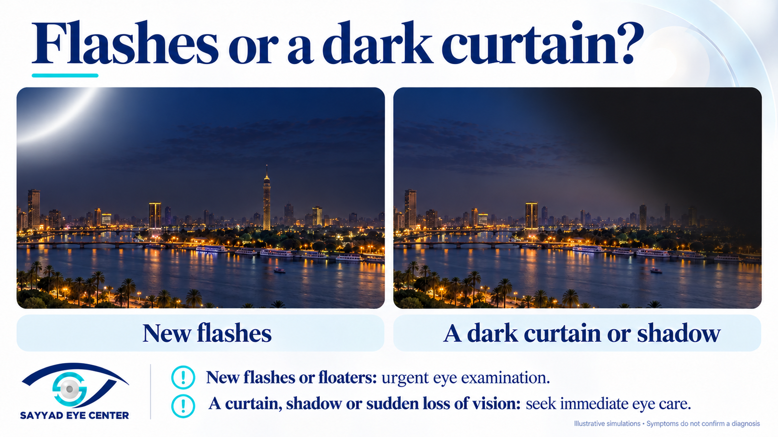
New flashes, new floaters or a sudden increase in floaters need an urgent eye examination, preferably the same day.
Seek immediate eye care if you notice:
A dark curtain or shadow spreading across your vision.
A missing area of side vision.
A sudden shower of dots or many new floaters.
A sudden reduction in vision.
Do not wait for pain. A retinal tear or retinal detachment can be painless.
These instructions still apply if you recently had a reassuring examination. A new change in symptoms needs a new assessment.
What is posterior vitreous detachment?
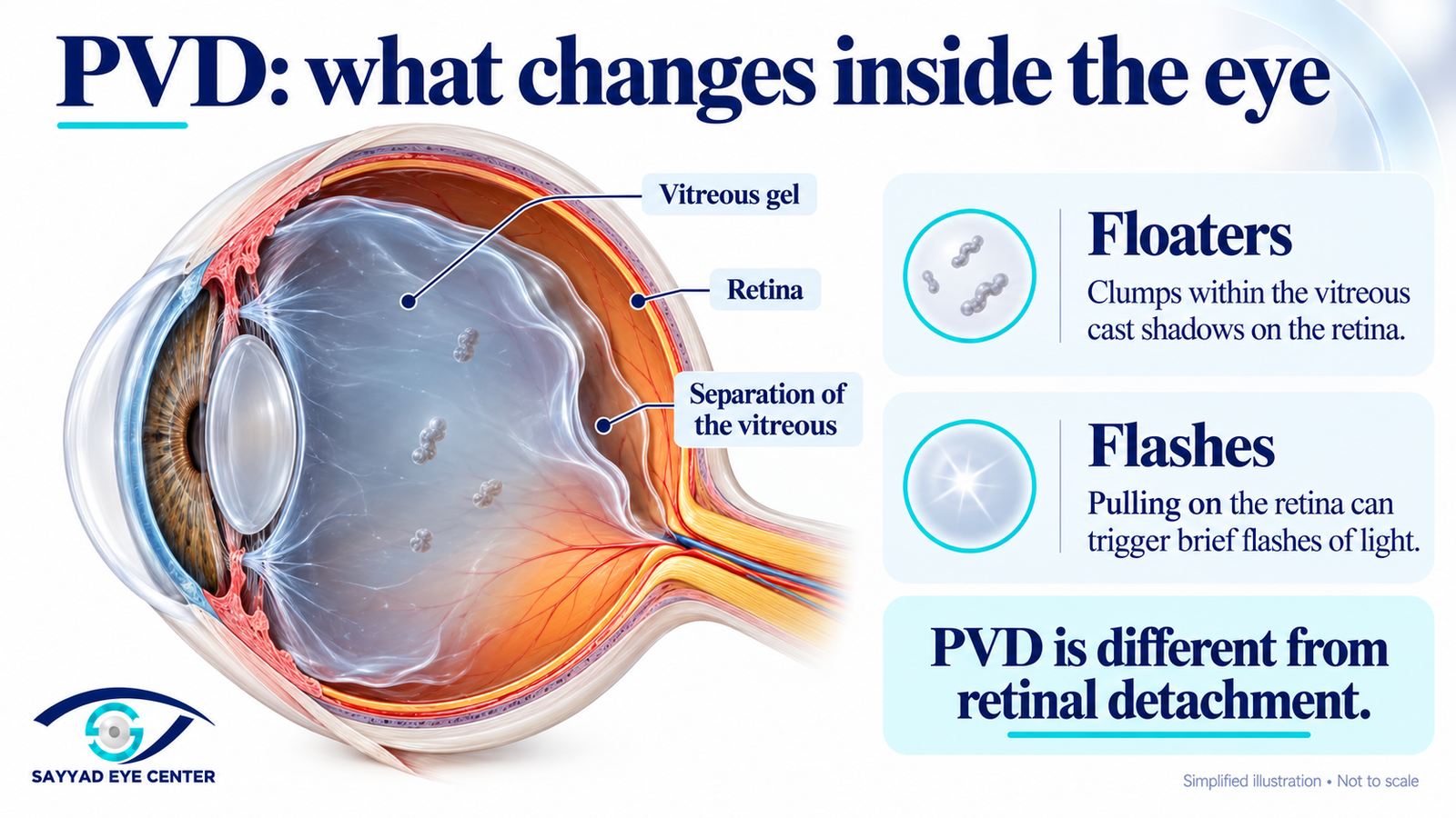
The vitreous is the clear gel that fills much of the inside of the eye. It lies against the retina, the light-sensitive tissue lining the back of the eye.
As we get older, the gel becomes more liquid and changes its structure. It can gradually separate from the retinal surface. This separation is called posterior vitreous detachment.
PVD becomes more common with age. It can happen earlier in people with short-sightedness, particularly higher prescriptions, and may also occur following an eye injury or eye surgery.
It is an internal change within the eye. It does not mean that the eye itself is becoming detached.
Is PVD the same as retinal detachment?
No. They are different conditions.
In an uncomplicated PVD, the vitreous gel separates while the retina remains in its normal position. Most people do not need an operation.
Occasionally, the gel pulls firmly on a point where it is attached and creates a retinal tear. Fluid can pass through that tear and lift the retina away from the underlying tissue, causing a retinal detachment.
This is why we examine new flashes and floaters promptly. The examination helps distinguish a common vitreous change from a problem that needs treatment.
What do flashes and floaters look like?
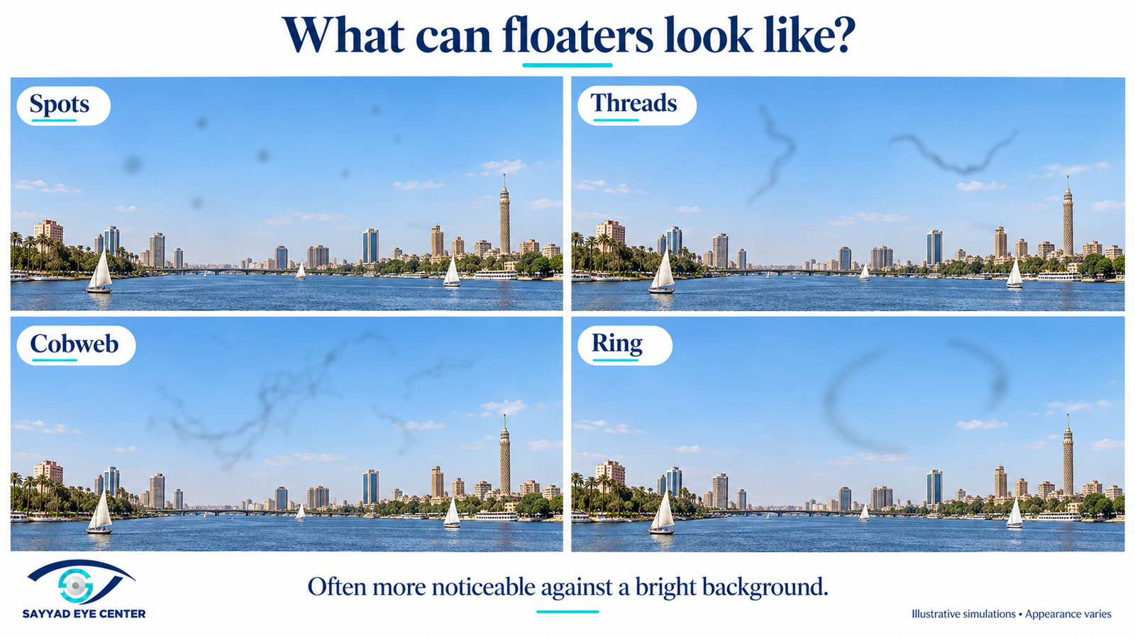
Floaters can resemble:
Small dots or specks.
Threads or strands.
Cobwebs.
A ring.
A translucent cloud that drifts across your vision.
They move as your eye moves and may continue drifting after you stop looking around. They are often more noticeable against a white wall, a bright sky or a computer screen.
Floaters cast shadows on the retina. They are inside the eye, so cleaning your glasses or washing the eye will not remove them.
PVD-related flashes are usually brief streaks or flickers, often at the side of your vision. They may be more noticeable in dim surroundings.
Not every flashing pattern comes from PVD. Migraine and other conditions can also cause visual symptoms, so a new or unfamiliar episode should not be diagnosed from its appearance alone.
What happens at your examination?
We ask when the symptoms started, which eye is affected and whether your vision has changed. Tell us about previous eye surgery, injury, retinal treatment or retinal detachment in either eye.
The assessment includes vision testing and a dilated retinal examination, including the outer edges of the retina.
Additional tests are sometimes helpful:
OCT: assesses the central retina and the relationship between the vitreous and the macula.
B-scan ultrasound: may be needed if the retina cannot be seen clearly, for example because of bleeding inside the eye.
A normal central OCT does not replace examination of the peripheral retina or rule out a tear at its edge.
Bring your glasses and arrange transport home. Dilating drops can temporarily blur your vision and make bright light uncomfortable.
Why might you need another examination?
The vitreous may continue separating after the first visit. A retinal tear can therefore develop later, even when none was visible initially.
A large American registry study supports at least one repeat examination within six weeks after an acute PVD, with earlier review for higher-risk eyes. A UK prospective study also found that some tears developed during follow-up.
The timing depends on the examination. Bleeding, areas of retinal thinning, or a history of retinal tears or detachment may mean closer follow-up is needed.
Keep your follow-up appointment, but do not wait for it if symptoms change. More floaters, worsening flashes, a curtain or reduced vision should bring you back sooner.
What can you do after an uncomplicated PVD?
Once the retina has been examined and no tear or other complication has been found, ordinary daily life can usually continue.
Reading, phones and computers
You can generally read, watch television and use your phone or computer. These activities may make floaters more noticeable because of the bright background.
Use comfortable lighting and adjust screen brightness. Take breaks if your eyes feel tired.
Walking and everyday activities
Walking, preparing meals and light daily activities are generally possible. Bed rest is not a routine treatment for uncomplicated PVD.
Exercise and heavy lifting
There is no strong evidence that everyone with uncomplicated PVD must stop exercising. Some specialists advise temporarily avoiding heavy lifting, high-impact exercise and contact sports during the early weeks, particularly when the eye has additional risk factors.
Restrictions should reflect the retinal findings. Advice may differ if there is bleeding, a tear or recent retinal treatment.
Driving
Do not drive while your vision is blurred after dilation or if floaters interfere with seeing safely. Resume when you can see clearly enough to drive safely and meet the applicable driving requirements.
How long do flashes and floaters last?
Flashes often settle as the vitreous separation progresses. Floaters commonly become less distracting over several weeks to months.
Some floaters remain visible, especially against a bright background. Improvement does not necessarily mean that every floater disappears. They may move away from the centre of vision, and you may gradually notice them less.
Persistent floaters can be frustrating even when the retina is healthy. Mention them at follow-up, particularly if they interfere with reading, work or driving.
A sudden new change after a period of improvement still needs reassessment.
Do eye drops or supplements help?
Eye drops, vitamins and eye exercises have not been established as treatments that reverse PVD or reliably remove its floaters.
Lubricating drops may help if you also have dry eyes, but they do not treat the vitreous change itself.
For most uncomplicated cases, the approach is examination, appropriate follow-up and allowing time for the symptoms to become less intrusive.
What if floaters continue to interfere with daily life?
Most people do not need a procedure. However, persistent floaters that significantly affect daily activities deserve a discussion with a retina specialist.
Vitrectomy, an operation that removes the vitreous gel, may be considered in selected cases. It can reduce floaters, but it also carries surgical risks, including cataract development, retinal tears or detachment, and infection.
Laser treatment for floaters is another option sometimes discussed. Its suitability depends on the floater and its position, and it should not be presented as a routine or risk-free cure.
The decision depends on how much the symptoms affect you, what the examination shows and whether the expected benefit justifies treatment.
Frequently asked questions
Does having PVD mean I will lose my sight?
An uncomplicated PVD usually does not cause permanent sight loss. The reason for urgent assessment is to identify the smaller group of patients who have an associated retinal tear, bleeding or detachment.
Once the retina has been checked, understanding the diagnosis and knowing the warning signs can make the symptoms much less worrying.
If I have a retinal tear, will I need a major operation?
Some retinal tears can be treated with laser or freezing treatment to reduce the risk of retinal detachment. A retinal detachment may require surgery.
Not every PVD needs laser. Treatment is based on the retinal findings.
Can PVD happen in the other eye?
Yes. The other eye may develop similar changes at a different time.
New symptoms in that eye need their own examination, even if the first eye had an uncomplicated PVD.
I have had the same floater for years. Is that an emergency?
A longstanding floater that has already been assessed and remains unchanged is different from the sudden appearance of new floaters.
Arrange an examination if it has never been checked. Seek urgent assessment if its appearance changes suddenly, more floaters develop, flashes appear or your vision is affected.
Can I still have a retinal tear if I can read normally?
Yes. A peripheral tear may not initially affect your central reading vision. Being able to read clearly does not replace a dilated retinal examination when symptoms are new.
Do I need an eye patch or a special sleeping position?
Neither is a routine treatment for uncomplicated PVD. Specific positioning instructions usually relate to another condition or a retinal procedure, rather than PVD itself.
Can I travel or fly?
Uncomplicated PVD itself does not usually prevent flying.
If symptoms are new, have the retina assessed before travelling and consider whether you will have access to urgent eye care and your planned follow-up.
If you have undergone retinal surgery and have a gas bubble inside the eye, do not fly until the gas has completely disappeared and the retinal surgeon has confirmed that flying is safe.
Why do I still see floaters after a normal examination?
A reassuring retinal examination means that no tear or detachment was found at that visit. It does not remove the vitreous changes that create the floaters.
The floaters can remain while gradually becoming less noticeable. A sudden increase or a change in vision needs another examination.
Can pineapple or pineapple juice help with floaters?
You may have heard that pineapple can help dissolve floaters. A pilot study published by Horng and colleagues in the Journal of American Science in 2019 reported improvement after participants ate pineapple daily for three months. However, the study had important limitations: it did not include an untreated or placebo comparison group, so it could not establish whether the improvement came from pineapple or from the natural changes that often occur with floaters. It studied pineapple pieces, not pineapple juice.
A later study by Ma and colleagues, published in the Journal of Clinical Medicine in 2022, reported improvement with supplements containing bromelain, papain and ficin—enzymes derived from fruit. This study tested enzyme capsules rather than pineapple juice, and its findings need confirmation in larger, independent studies.
The research is interesting, but the evidence remains limited. It does not establish that pineapple juice dissolves floaters or treats PVD. New floaters or flashes still need a retinal examination.
Sources: Horng et al., 2019 — pineapple pilot study; Ma et al., 2022 — fruit-enzyme supplement study.
Before you leave the clinic
Make sure you know:
Whether a retinal tear or another complication was found.
When your next examination is due.
Whether any activity restrictions apply to your eye.
Where to seek urgent care if symptoms change.
The most important instruction is simple: new or worsening flashes, a sudden increase in floaters, a curtain or reduced vision should not wait for a routine appointment.
Related article
Retinal detachment: warning signs, treatment and recovery
Contact Sayyad Eye Center
For an appointment or follow-up, contact Sayyad Eye Center on +20 100 008 8168. Confirm whether your visit is in Mohandeseen or New Cairo, and bring any previous eye reports.
If you develop urgent warning symptoms and the centre is unavailable, attend an eye emergency service. Do not wait for a message reply.
Medical review
Medically reviewed by Dr Fouad El Sayyad
American Board certified ophthalmologist
Fellowship in Cornea, External Eye Disease and Refractive Surgery
Washington University in St Louis, USA
References
American Academy of Ophthalmology. Kim SJ, Bailey ST, Kovach JL, et al. Posterior Vitreous Detachment, Retinal Breaks, and Lattice Degeneration Preferred Practice Pattern. Ophthalmology. 2025;132(4):P163–P196. PubMed.
American Society of Retina Specialists. Posterior Vitreous Detachment.
Moorfields Eye Hospital NHS Foundation Trust. Flashes and floaters.
Royal National Institute of Blind People. Posterior vitreous detachment.
Nixon TRW, Davie RL, Snead MP. Posterior vitreous detachment and retinal tear—a prospective study of community referrals. Eye. 2024;38(4):786–791. PubMed.
Vangipuram G, Li C, Li S, et al. Timing of Delayed Retinal Pathology in Patients Presenting with Acute Posterior Vitreous Detachment in the IRIS Registry (Intelligent Research in Sight). Ophthalmology Retina. 2023;7(8):713–720. PubMed.
American Society of Retina Specialists. Vitrectomy for Floaters.