
Retinal Detachment
New flashes, floaters or a curtain in vision need urgent assessment. Learn about retinal detachment treatment, surgery, recovery and gas-bubble precautions.
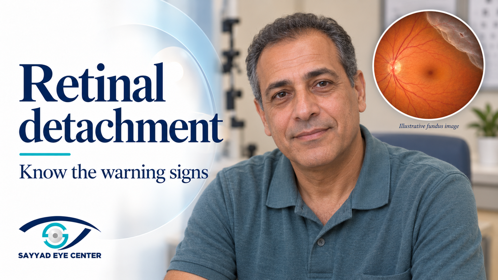
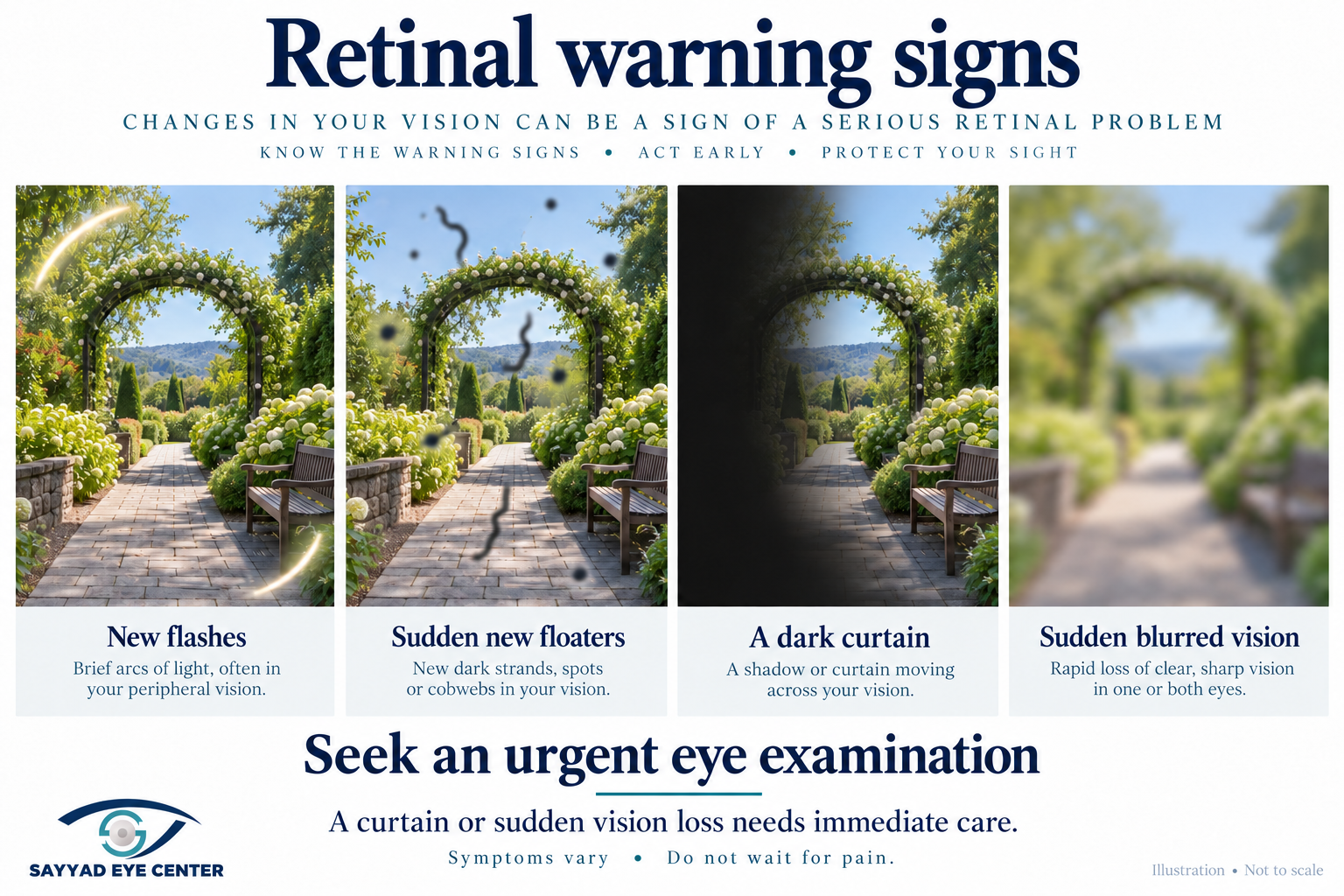
New flashes, a sudden shower of floaters, a dark curtain or sudden loss of vision need an urgent eye examination. A curtain or loss of vision means seek care immediately. Do not wait for pain or for an online appointment reply. If prompt assessment is unavailable at the center, go to the nearest emergency eye service.
Retinal detachment is treatable, and prompt care offers the best opportunity to protect sight. This guide explains what the symptoms mean, how a retinal tear differs from a detachment, and what to expect from treatment. It also covers recovery and the important restrictions that apply when an air or gas bubble is used inside the eye.
What is retinal detachment
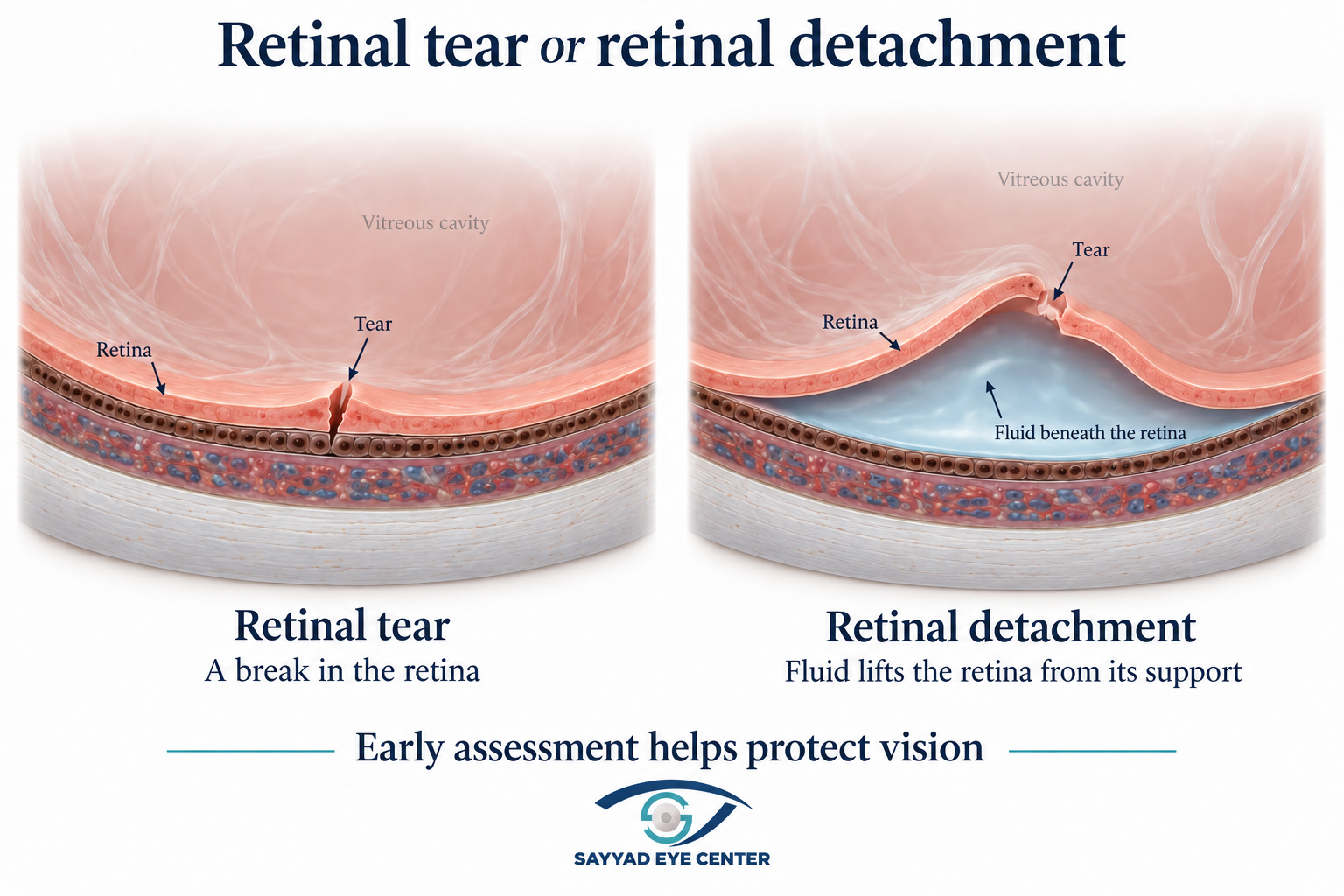
The retina is the light-sensitive tissue lining the back of the eye. A detachment occurs when it lifts away from the supporting tissue beneath it. In the most common form, fluid passes through a tear or hole and collects underneath. The separated retina cannot work normally, and untreated detachment can cause permanent loss of vision.
Symptoms to recognise
• Many new floaters, which may look like dots, strands or cobwebs.
• New flashes of light, often toward the side of your vision.
• A dark shadow or curtain covering part of the view.
• Sudden blurred vision, missing side vision or loss of central detail.
Retinal detachment is usually painless, and the eye can look normal from the outside. You do not need to have all these symptoms. Even if flashes or floaters settle, an urgent assessment is still needed when they are new or suddenly increase.
A vitreous detachment is not the same as a detached retina
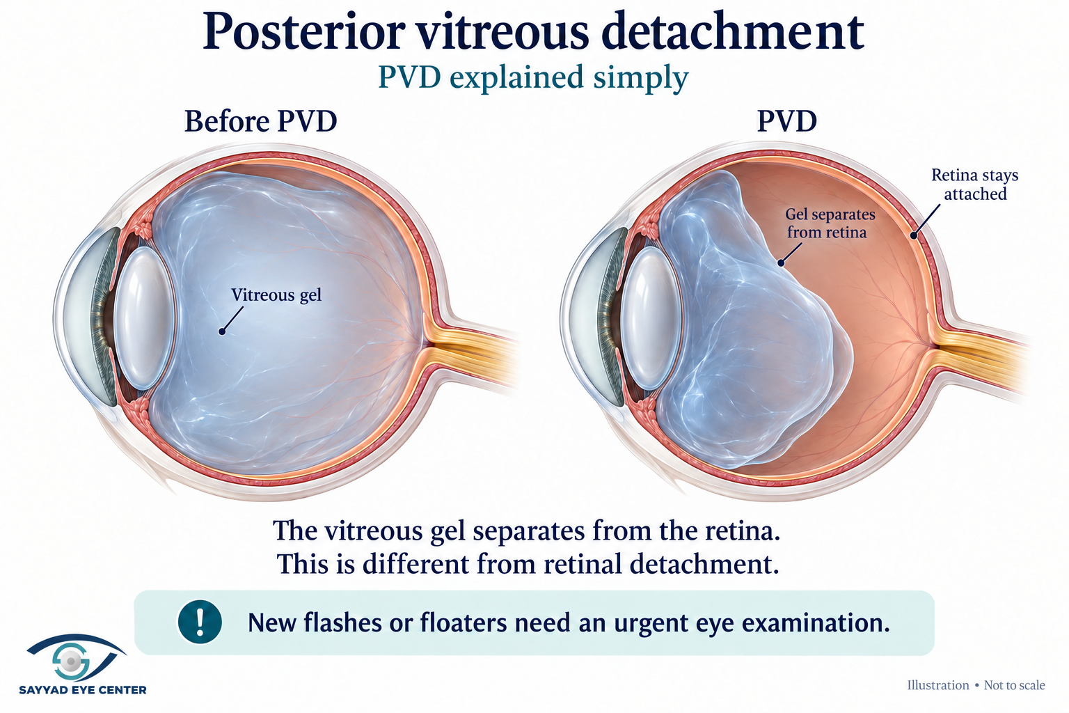
The vitreous is the clear gel inside the eye. As it changes with age, it can separate from the retina; this is called posterior vitreous detachment, or PVD. Most PVDs do not damage the retina. Sometimes the gel pulls hard enough to cause a tear, which may then lead to a retinal detachment. Symptoms alone cannot reliably distinguish these conditions.
Who is at higher risk
Risk factors include high short-sightedness, increasing age, eye injury, a previous retinal tear or detachment, certain weak areas in the peripheral retina and family history. Previous eye surgery, including cataract surgery, can also increase risk in some eyes. Advanced diabetic eye disease can cause a different form of detachment through scar tissue pulling on the retina.
Correcting a high myopia prescription with glasses, laser or an implantable lens does not remove the retinal risk associated with a long eye. Continue the retinal checks your doctor recommends.
Why the macula matters
The macula is the central retina used for reading and recognising faces. If it is still attached, treatment aims to protect that central vision. If it has detached, surgery can still help, but recovery depends partly on how long it has been affected and the condition of the retina. Both situations need prompt specialist assessment; do not assume that a delay is safe because central vision is still clear or has already fallen.
Diagnosis and treatment options
What happens at the examination
The doctor uses dilating drops to examine the retina, including its far edges, for tears and detachment. OCT can show whether the central retina is affected, but does not replace a peripheral retinal examination. Ultrasound helps when blood, a dense cataract or another opacity prevents a clear view. Bring previous reports and a medicine list, and arrange transport because dilation may blur vision temporarily.
Different types need different treatment
Rhegmatogenous detachment is caused by a tear or hole. Tractional detachment occurs when scar tissue pulls the retina away, often in advanced diabetic retinopathy. Exudative or serous detachment is caused by fluid leakage without a tear, for example with inflammation or other underlying disease. Treating the cause is essential; not every type is managed with the same operation.
Treating a retinal tear before a larger detachment develops
Laser or freezing treatment called cryotherapy can create an adhesion around a suitable tear, reducing the chance of fluid spreading underneath the retina. Not every small hole or area of peripheral thinning needs treatment; the retina specialist decides according to its features and your risk. Treatment reduces risk but does not prevent every future tear, so new symptoms still need urgent review.
Surgery for a detached retina
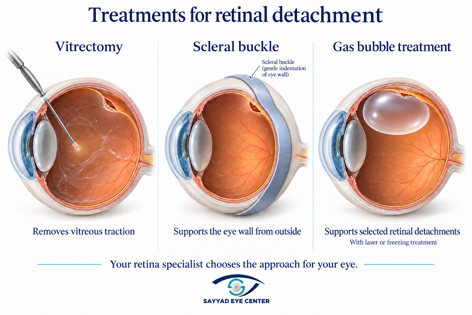
The choice depends on the tears, extent of detachment, condition of the lens and vitreous, and whether scar tissue is present. Procedures may be combined. Your surgeon will explain the anaesthesia and the plan for your eye.
Vitrectomy
How it helps: Removes vitreous traction. Tears are treated and air, gas or silicone oil may support the retina.
What matters afterward: Drops, reviews and sometimes a prescribed head position. Gas affects travel.
Scleral buckle
How it helps: A silicone band or segment on the outside of the eye supports the area of the break and reduces traction.
What matters afterward: The buckle usually stays in place. Glasses prescription may change.
Pneumatic retinopexy
How it helps: A gas bubble supports selected detachments, with laser or freezing to seal the break.
What matters afterward: Suitable only for selected cases; precise positioning and close reviews are important.
Laser alone is usually not enough for an established retinal detachment. A retina specialist may use it for carefully selected small, limited detachments; this is not a reason to delay assessment or substitute routine laser for recommended surgery.
Benefits and realistic expectations
The aim is to reattach the retina, preserve sight and allow as much visual recovery as the eye can achieve. Many detachments can be repaired successfully, although some need more than one procedure. An attached retina does not guarantee that vision returns fully to its previous level, particularly if the macula was affected.
Risks to discuss calmly
Like any surgery, retinal surgery carries risks. These include infection, bleeding, pressure changes, cataract progression, persistent visual changes and further detachment. Scar tissue, known as proliferative vitreoretinopathy, can pull on the retina again and require additional surgery. Risks vary considerably with the case; they should be weighed against the risk to sight from leaving the detachment untreated.
Recovery and frequently asked questions
Your recovery plan
Use the prescribed drops and attend early and later reviews to check the retina and eye pressure. Follow your written instructions for sleep, exercise, lifting, work and driving. Some patients need a particular head position to place the bubble against the treated area; it is not automatically face down for everyone. Ask for a demonstration and discuss any neck or mobility difficulty before leaving.
Vision may be very blurred while a gas bubble remains, and improvement can continue over weeks or months. Some blur or distortion may remain despite successful repair. Mild discomfort may occur, but increasing pain, marked redness, worsening vision or a new curtain needs urgent contact with the surgical team or an emergency eye service.
Air or gas inside the eye
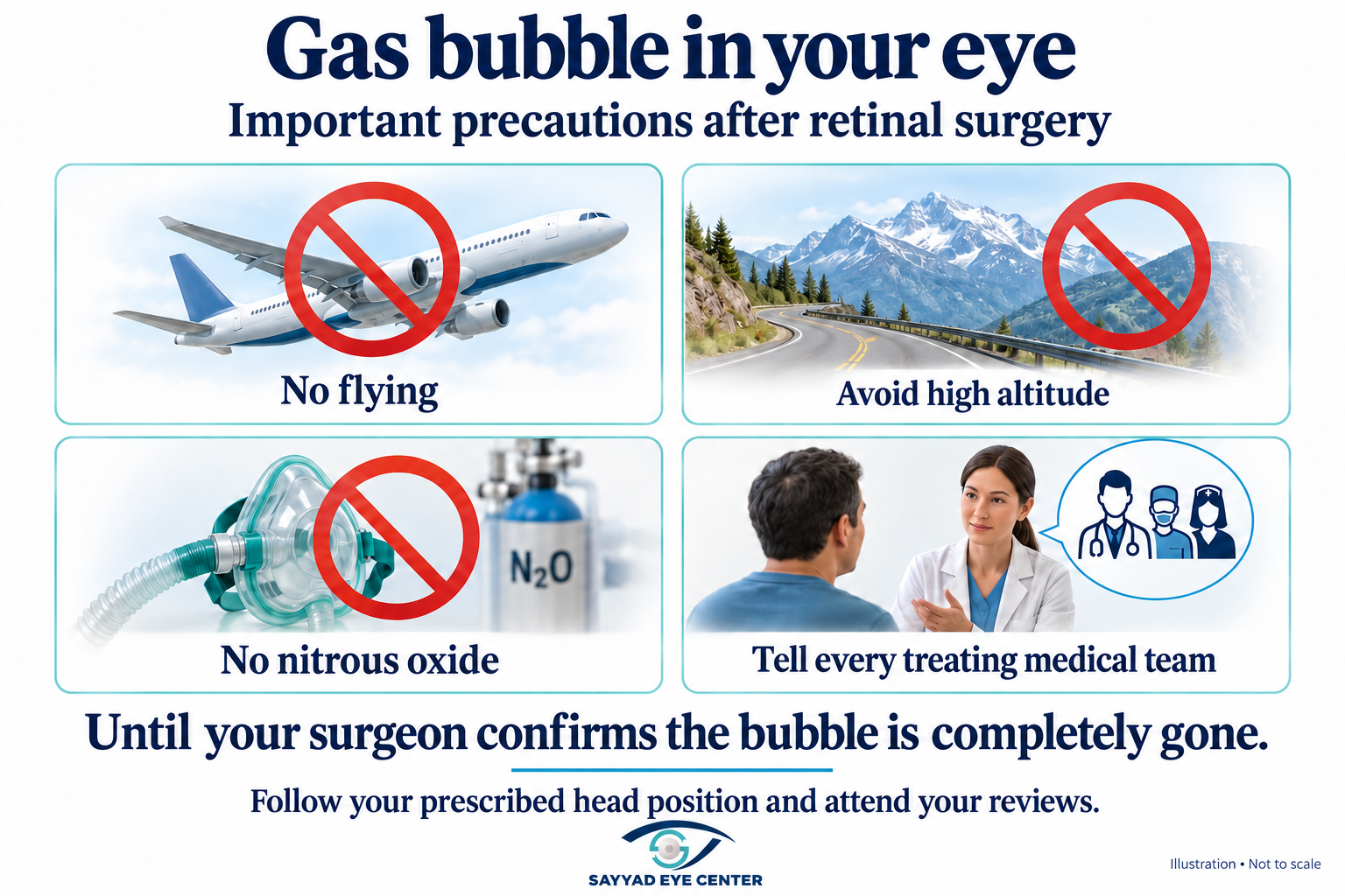
Do not fly, travel to high altitude or scuba dive while an air or gas bubble remains. Fly only after the surgeon confirms the bubble has completely gone and clears you to travel. Do not decide from the calendar or because the bubble seems small. Tell every treating medical or dental team that you have gas in the eye: nitrous oxide, sometimes used for anaesthesia or pain relief, must be avoided while the bubble remains. Keep any gas-alert card supplied by the clinic.
Patients travelling from abroad
If symptoms start in the UAE, Saudi Arabia, another GCC country or Europe, seek urgent care where you are rather than delaying assessment to travel to Egypt. For planned care or follow-up at Sayyad Eye Center, share your reports and discuss the likely length of stay before booking. Gas may prevent a flight home for weeks. Silicone oil is different from gas, but does not automatically make early travel appropriate; your surgeon must confirm the full plan.
Frequently asked questions
Do floaters always mean retinal detachment
No. Longstanding stable floaters are common. New floaters or a sudden increase, especially with flashes or a shadow, need urgent examination.
Can drops repair a retinal detachment
Drops cannot repair the usual tear-related detachment. Some fluid-related detachments need treatment of an underlying disease, which may include medicine.
How long does gas stay in the eye
It depends on the gas and its concentration. It may remain for weeks. Follow the surgeon’s clearance, not an estimated disappearance date.
Does silicone oil disappear by itself
No. It may need a separate removal procedure after the retina is stable; some complex eyes need it for longer.
Can detachment happen again or affect the other eye
Yes. New tears or scar tissue can lead to another detachment. The other eye also needs assessment and the follow-up advised for its risk.
Does everyone regain full vision
No. The aim is the best achievable sight for that eye. Macular involvement, duration, complexity and other eye disease affect the result.
Contact Sayyad Eye Center in New Cairo or Mohandeseen to arrange retinal assessment and confirm specialist availability. Patients from Sheikh Zayed or 6th of October should confirm the appropriate branch. For warning symptoms, obtain urgent local care if a prompt center appointment is unavailable.
References
Medical reviewer: Dr Reem Osama, Consultant Vitreoretinal Surgeon; Fellow of the Royal College of Surgeons of Edinburgh in Ophthalmology; Lecturer at Ain Shams University, Cairo, Egypt.
المراجع ومصادر المعلومات
American Academy of Ophthalmology sources for patient education and clinical review. References are collected at the end of the article in each published language. No private-clinic or wiki references are used. Updated 6 October 2026.
AAO — Detached Retina https://aao.org/eye-health/diseases/detached-torn-retina
AAO — Vitrectomy patient guide https://store.aao.org/media/resources/051220/051220-vitrectomy-rf1.pdf
AAO — Posterior Vitreous Detachment https://www.aao.org/eye-health/diseases/what-is-posterior-vitreous-detachment
AAO — Posterior Vitreous Detachment Retinal Breaks and Lattice Degeneration Preferred Practice Pattern https://www.aaojournal.org/article/S0161-6420%2824%2900787-5/fulltext
This article is a patient education draft for medical approval. Dr Reem Osama’s final medical approval is required before publication. The treating retina specialist determines treatment and recovery instructions. Do not use this guide to postpone urgent examination.
هذا النص مسودة تثقيفية للاعتماد الطبي قبل النشر، وتلزم موافقة د. ريم أسامة النهائية قبل النشر. يحدد الاختصاصي المعالج العلاج وتعليمات التعافي. لا تستخدم الدليل لتأجيل الفحص العاجل.